

Master your cellular energy production for peak performance, vitality, and longevity, Start the 11-part series below.
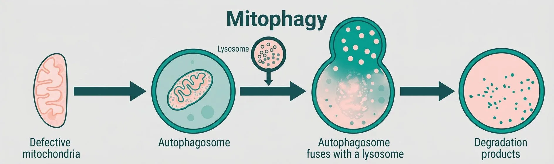
Mitochondria do not last forever; they have a half life of 28 days. In addition, they accumulate damage over time, become less efficient and dysfunctional, and can begin to cause harm if they are not removed. The cellular process that identifies and removes damaged mitochondria, is called mitophagy.
Mitophagy is one of the most important but least appreciated aspects of mitochondrial health. It is not merely a cleanup mechanism. It is a quality control system on which the integrity of the entire mitochondrial population depends. Without it, damaged organelles accumulate, energy production falters, and the cell becomes increasingly dysfunctional.
Mitophagy is a specialized form of autophagy (from the Greek word for self-eating), the broader process by which cells break down and recycle their own components. While general autophagy can target a wide range of cellular material, mitophagy specifically targets mitochondria that have been identified as damaged, dysfunctional, or no longer needed.
The word describes the selective degradation of mitochondria, a process that is distinct from general cellular recycling in both its triggers and its precision. Mitophagy is not random. It is a targeted process that requires the damaged mitochondrion to be flagged, recognized, and removed before they begin to cause dysfunction.
The process of mitophagy involves several coordinated steps.
Mitochondria are dynamic structures that continuously divide (fission) and merge (fusion) in response to cellular conditions. They are often connected to one another, forming dynamic, tube-like structures in the cell known as the mitochondrial network. Fission serves a critical quality control function: it allows the cell to isolate damaged parts of a mitochondrion or damaged segments of the mitochondrial network into smaller, separate units.
When a portion of the mitochondria or mitochondrial network is damaged, fission separates it from the healthy network. This isolation prevents the damaged segment from rejoining the healthy population through fusion. The separated, damaged mitochondrion is then available for identification and removal by absorption by the mitochondria itself.
The most well-characterized mechanism of mitophagy involves two proteins: PINK1 (PTEN-induced kinase 1) and Parkin, a ubiquitin ligase. Under normal conditions, PINK1 is rapidly degraded inside healthy mitochondria and does not accumulate. When a mitochondrion is damaged and its membrane potential is lost, PINK1 can no longer be processed and instead accumulates on the outer mitochondrial membrane.
This accumulation of PINK1 attracts Parkin, which attaches ubiquitin tags to proteins on the outer mitochondrial membrane. These tags act as molecular flags: this mitochondrion is damaged and should be removed. Autophagy receptors recognize these flags and direct the formation of an autophagosome, a double-membrane vesicle that engulfs the tagged mitochondrion. The autophagosome then fuses with a lysosome, where the mitochondrion is broken down and its components are recycled or eliminated.
The components released through mitophagy, amino acids, lipids, and other molecular building blocks, are returned to the cell’s general pool of materials and can be reused. Mitophagy is not simply removal. It is recycling, a way of recovering valuable resources from structures that can no longer serve their intended function.
The importance of mitophagy becomes clear when you consider what happens when damaged mitochondria are not removed.
Excessive reactive oxygen species (ROS) production. Damaged mitochondria with compromised electron transport chains generate disproportionate amounts of reactive oxygen species, highly reactive molecules that damage proteins, lipids, and DNA. A healthy cell manages ROS through antioxidant defenses, but damaged mitochondria can produce ROS at levels that overwhelm these defenses.
Reduced ATP production. Damaged mitochondria produce less ATP per unit of fuel consumed. As the ratio of damaged mitochondria in the cell’s mitochondrial population increases, overall energy production falls thus starting a downward bioenergetics spiral which impairs all biological functions that depend on it.
Amplification of inflammation. Damaged mitochondria release signals that activate inflammatory pathways. This can trigger chronic low-grade inflammation that damages surrounding tissues and contributes to disease conditions ranging from metabolic disease to neurodegeneration.
The ROS vicious cycle. Damaged mitochondria produce more ROS, which damages more mitochondria, which produce more ROS. Without mitophagy to interrupt this cycle, the dysfunction continues endlessly.
Mitophagy and mitochondrial biogenesis are not independent processes. They are two halves of a continuous renewal cycle. Biogenesis creates new, functional mitochondria. Mitophagy removes old, damaged ones.
When both processes are operating well, the mitochondrial population is continuously refreshed, maintaining high average quality and high overall energy output leading to an upward spiral in bioenergetics. When either process is impaired, the balance shifts.
If biogenesis slows without a corresponding slowdown in mitophagy, the cell may clear mitochondria faster than it replaces them. If mitophagy is impaired while biogenesis continues, damaged mitochondria accumulate alongside new ones, degrading overall quality and increasing oxidative stress. The optimal state is one in which both processes are active and balanced.
The connection between impaired mitophagy and disease is well documented by researchers in the last 40 years:
Parkinson’s disease. Mutations in the genes encoding PINK1 and Parkin are among the most common genetic causes of Parkinson’s disease. Defective mitophagy leads to accumulation of damaged mitochondria in neurons, contributing to the cell death patterns characteristic of the disease.
Aging. Mitophagy efficiency declines with age. The same decade-by-decade decline in mitochondrial density that characterizes normal aging is accompanied by a reduced capacity to clear damaged mitochondria. The two processes reinforce each other leading to a downward spiral in bioenergetics.
Metabolic disorders. Impaired mitophagy in liver and muscle cells has been linked to insulin resistance and the metabolic dysfunction associated with obesity and type 2 diabetes.
Neurodegenerative conditions. Beyond Parkinson’s, impaired mitophagy has been implicated in Alzheimer’s disease and other neurodegenerative conditions in which neuronal energy deficits and oxidative damage play central roles.
The factors that support mitophagy overlap considerably with those that support mitochondrial health broadly:
Exercise. Physical activity induces mitophagy as well as biogenesis, stimulating the removal of damaged mitochondria while simultaneously signaling the production of new ones. This dual effect makes exercise particularly powerful as a mitochondrial health intervention.
Caloric restriction and fasting. Periods of reduced caloric intake activate autophagy broadly, including mitophagy, through AMPK and mTOR signaling pathways.
Quality sleep. Cellular cleanup processes, including mitophagy, are most active during sleep. Disrupted sleep impairs these processes and allows damaged cellular components to accumulate.
Avoiding mitochondrial damage. Reducing exposure to ultra-processed foods, high fructose corn syrup, environmental toxins,
excessive alcohol, and chronic psychological stress, reduces the burden that mitophagy must manage.
When mitophagy is running efficiently and in balance with biogenesis, the cell maintains a healthy, high-quality mitochondrial population. When it is not, the compounding effects of accumulated damage drive the cellular dysfunction associated with aging and disease.
Mitochondrial health is a rich, interconnected field. The pages in this knowledge hub explore each dimension in greater depth, from the molecular mechanics of biogenesis and mitophagy to the relationship between mitochondria and specific health conditions.
This content is for educational purposes only and is not medical advice. Consult a healthcare professional for guidance specific to your situation.
Studies, articles and talks behind this page.
Youle, Richard J., and Derek P. Narendra. “Mechanisms of Mitophagy.” Nature Reviews Molecular Cell Biology 12, no. 1 (2011): 9–14. https://doi.org/10.1038/nrm3028. Free full text
Narendra, Derek P. “Managing Risky Assets – Mitophagy in Vivo.” Journal of Cell Science 134, no. 19 (2021): jcs240465. https://doi.org/10.1242/jcs.240465. Free full text
Fang, Evandro F., Yujun Hou, Konstantinos Palikaras, Bryan A. Adriaanse, Jesse S. Kerr, Beimeng Yang, Sofie Lautrup, et al. “Mitophagy Inhibits Amyloid-β and Tau Pathology and Reverses Cognitive Deficits in Models of Alzheimer’s Disease.” Nature Neuroscience 22, no. 3 (2019): 401–12. https://doi.org/10.1038/s41593-018-0332-9. Free full text
BlueOakNx. “Salugenesis: How Cells Adapt to Stress.” BlueOakNx Blog.
BlueOakNx. “Mitochondria 101.” BlueOakNx Science.
This content is for educational purposes only and is not medical advice. Consult a healthcare professional for guidance specific to your situation.