
Role of Mitochondrial Dysfunction in Autism Spectrum Disorders
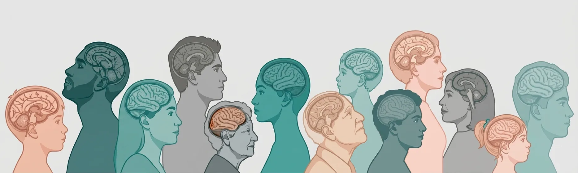
More than 80 years have passed since Dr. Kanner first diagnosed autism; initially, it was referred to as the Kanner Syndrome. Today, as per the U.S. Centers for Disease Control, better diagnosis and increased awareness has resulted in identifying approximately 1 in 31 children (aged 8 years) with the Autism Spectrum Disorder (ASD).
ASD occurs in all socioeconomic, racial, and ethnic groups, with the diagnosis being three times more common in boys than in girls. The symptoms range from problems with social interaction and repetitive behavior to delayed speech, delayed cognition, sensory sensitivities, emotional dysregulation, and more.
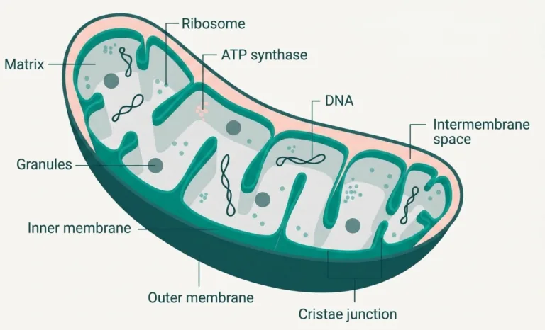
Since the first diagnosis of autism, many causes have been proposed to explain this challenging disorder, ranging from genetics and pollution to vaccines. In this article, we focus on more recent research that finds a connection between dysfunctional mitochondria (the key organelle responsible for bioenergetics and life) and ASD pathophysiology.
Connection between Dysfunctional Mitochondria and Autism Spectrum Disorder
Since the beginning, the diagnosis of ASD was based on behavioral observations and genetic architecture; however, evidence suggests that individuals with ASD exhibit measurable mitochondrial abnormalities, requiring a shift in the assessment paradigm.
This shift positions mitochondrial dysfunction not as a consequential comorbidity, but as a key driver of ASD pathophysiology.
The brain’s high metabolic demand makes it especially vulnerable to mitochondrial impairment. The brain (only 2% of the body’s weight) uses approximately 20 percent of the body’s total energy, making it uniquely sensitive to the Cellular Bioenergetic Threshold, at which a cell’s energy demand exceeds its mitochondrial supply (reserve capacity), leading to dysfunction.
When mitochondrial capacity falls below this threshold, the resulting energy rationing manifests as the core deficits and behavioral challenges associated with autism (Naviaux 2014).
How Metabolic Deficits Lead to Neurodevelopmental Disorders
According to Robert Naviaux, MD, PhD, mitochondria are the primary initiators of the Cell Danger Response (CDR) – an evolutionary defense mechanism in which mitochondria shift their focus from growth and repair to survival.
The CDR is a protective, short-term mechanism to heal, but when it gets stuck in a chronic state and is unable to complete the healing cycle, it contributes to conditions such as autism and other neurodevelopmental disorders.
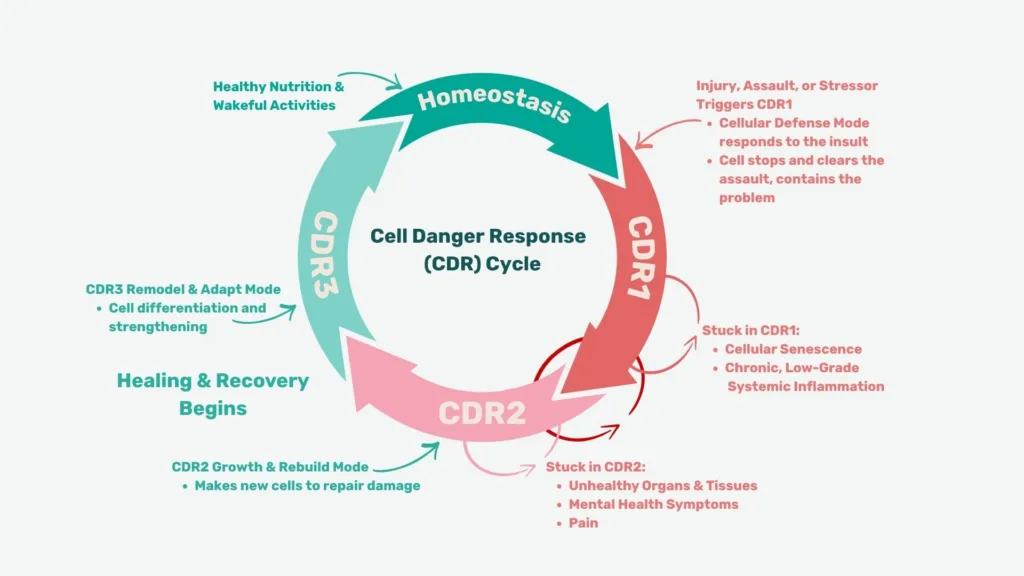
The mitochondria remain in a defensive, glycolytic state rather than an oxidative, energy-producing or repair state. This dysfunction prevents cells from reaching the bioenergetic threshold required for high-energy-demand neurodevelopmental processes, such as synaptic pruning, myelination, and the maintenance of complex neurotransmitter pathways (Goh et al. 2014).
Neurotransmission, Energy Rationing, and Behavioral Manifestations
When cellular energy is diminished, the brain prioritizes survival over other functions, such as social cognition and impulse control, affecting neurotransmitters responsible for behavioral stability, including sleep, impulse control, self-harm, and sensory overload.
Each of these is described briefly in the following subsections.
Sleep and Circadian Rhythm
Dysfunctional mitochondria can fail to power deep, restorative sleep. Reduced bioenergetics can affect melatonin levels and the regulation of GABAergic signaling.
This reduction can lead to chronic insomnia, frequently observed in ASD, which can further deplete cellular bioenergetic threshold (Adams et al. 2021).
Impulse Control and Executive Function
With reduced bioenergetics and ATP levels, the brain deprioritizes prefrontal cortex (PFC) function and shifts control to the primitive limbic system, which has a lower energy demand.
This issue manifests clinically as poor impulse control and hyperactivity (Rose et al. 2018).
Self-Harm and Sensory Overload
Dysfunctional mitochondria can and do produce excessive reactive oxygen species (ROS), thereby enhancing cellular oxidative stress and neuroinflammation and affecting sensory processing.
This issue, combined with reduced bioenergetic levels, can lead to self-injurious behaviors (SIB), a consequence of the overall metabolic exhaustion. When the brain cannot afford the energy to process sensory inputs, the system enters a state of catastrophic overwhelm (Rossignol and Frye 2012).
Social Engagement and Academic Performance
It is important to understand the connection between internal energy and external behavior.
Consider the brain similar to a smartphone, and mitochondria are the battery. Socializing, learning, and regulating emotions are like energy-consuming apps that run constantly in the background of all individuals.
At a 90% charged state, the mitochondrial battery can support the body’s energy demands. At a 10% charged state, the cell shifts to Power Save Mode. Continuing the phone analogy, in this situation, the screen dims, the CPU slows down, and non-essential functions turn off.
Social interaction is one of the most metabolically demanding tasks the human brain performs, requiring rapid integration of verbal, nonverbal, and emotional information.
For individuals with ASD, who often operate within a compromised bioenergetic system, this demand is particularly challenging. When someone is near their bioenergetic threshold, the extra metabolic effort needed for social engagement can cause a cellular brownout, leading to withdrawal, sensory meltdowns, or complete shutdown.
Increasing the bioenergetic threshold through targeted mitochondrial support — improving mitochondrial quality, quantity, and function — can help build the adaptive reserves needed for complex social and academic processing.
This bioenergetic enhancement enables the brain to move away from a dysfunctional CDR and enter a physiological state better suited for growth, connection, and social interaction.
Benefits of Mitochondrial Restoration in ASD
Systemic support for ASD requires a shift from receptor-based pharmacology toward the restoration of cellular bioenergetics.
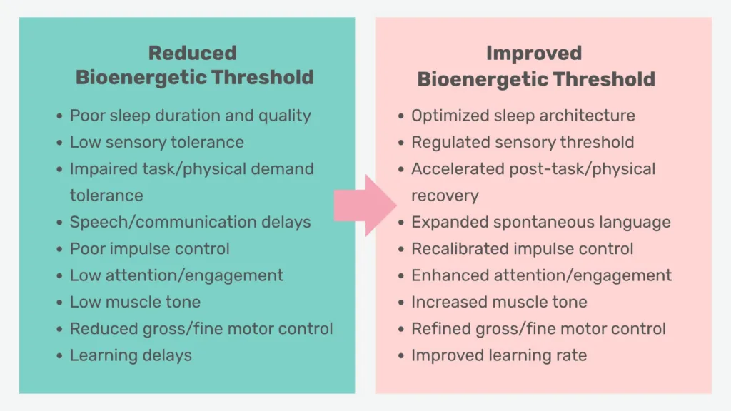
The table below lists, on the left, the typical results of a reduced bioenergetic threshold, while the right side lists the benefits of an improved bioenergetics threshold.
By optimizing cellular function at the mitochondrial level, there is a strong potential to improve the foundational energy that neuronal systems need to self-regulate and reach their full potential.
Read / Listen Next
A talk by Robert Naviaux, MD, PhD., on mitochondria and the emerging science of salugenesis
Sundeep Dugar, PhD., and Robert Lustig, MD, PhD., talk about why the brain’s energy crisis might be at the root of mental health challenges
References
- Adams, James B., Tapan Audhya, Elizabeth Geis, Robert J. Geier, Joan Feldman, Edward L. Hammer, Rosanne Coleman, and David W. Quig. 2021. “Biomarkers of Metabolic Control in Children with Autism Spectrum Disorders.” Frontiers in Physiology 12: 651–66. https://doi.org/10.3389/fphys.2021.643321.
- Goh, S., Dong, Z., Zhang, Y., DiMauro, S., & Peterson, B. S. (2014). Mitochondrial dysfunction as a neurobiological subtype of autism spectrum disorder: Evidence from brain imaging. JAMA Psychiatry, 71(6), 665–671. https://doi.org/10.1001/jamapsychiatry.2014.179.
- Naviaux, R. K. (2014). Metabolic features of the cell danger response. Mitochondrion, 16, 7–17. https://doi.org/10.1016/j.mito.2013.08.006.
- Rose, S., Niyazov, D.M., Rossignol, D.A. et al. Clinical and Molecular Characteristics of Mitochondrial Dysfunction in Autism Spectrum Disorder. Mol Diagn Ther 22, 571–593 (2018). https://doi.org/10.1007/s40291-018-0352
- Rossignol, D., Frye, R. Mitochondrial dysfunction in autism spectrum disorders: a systematic review and meta-analysis. Mol Psychiatry 17, 290–314 (2012). https://doi.org/10.1038/mp.2010.136.